|
|
|
A COLOUR ECHOTOMOGRAPHER WITH 4 - 32 COLOUR GRADATIONS FOR GETTING
A HISTOLOGICAL IMAGE IN VIVO AND WITH INTELECTUAL DIAGNOSTICS
PROVIDED
A colour echotomographer allows to estimate acoustic
density of a tissue by the means of colour coding of an echoimage.
This gives possibility to substantially enhance the image accuracy,
informative function and reliability. The device functions as an
ultrasound non-invasive microscope in vivo and displays a histological
image with colour gradations 4 up to 32.
The main device advantage is the possibility to get colour image
of organs and systems of different tissue densities.
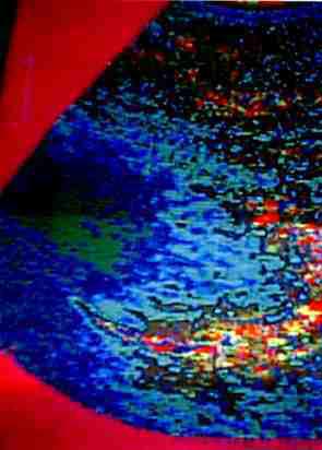
Heart valve
To date only the colour echotomographer in multicolour mode makes
it possible to greatly increase the image resolution.
Using a colour echotomographer we can distinguish different tissue
types, areas with intensive or poor circulation (ishemia, arterial
and venous hyperemia), unambiguously objectivate and thoroughly
interprete the presence of organ tissues of different density (tumours,
cirrhosis, postinfarct scares, oedemas, renal calculi and renal
sand) that are poorly and subjectively distinguished at the black&white
image, and the level of their microcirculation.
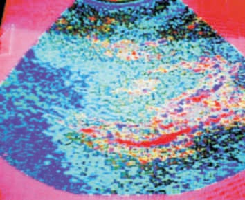
Muscle tissue with arteriolas (red-coloured)
Colour gradations in M-mode allows to investigate the heart valve
functionning as well as diseased tissue movement and functionning.
The use of colour echotomographer allows to avoid some imperfections
featuring up-to-date ultrasound systems:
- black&white ultrasound system without Dopplerogrephy gives no
possibility to get an informative image for clinical interpretation.
The doctor can make only subjective conclusions and miss the early
pathology not because of his ignorance but as a result of low
resolution of the even the best ultrasound systems;
- using an up-to-date ultrasound system with colour coding and
Dopplerography a doctor will get more information. But only the
circulation condition in major veins and arteries can be estimated,
not the microcirculation;
- a modern ultrasound system with energetic Doppler colour coding
allows us to get a one-colour picture of circulation in organs,
but there is no possibility to analize the tissue type in an organ,
particularly the areas with intensive circulation and to differentiate
arterial and venous discirculation that is substantial for an
individual pathogenetically grounded approach to treatment tactics;
- there is no ultrasound system giving a non-invasive contrasting
colour image of organs and/or systems allowing to objectively
diagnose tissues pathologies and their microcirculation level;
- a subjective interpretation of more or less echogenic organ
parts should not be used for objective control of the disease
development as it is not reliable at obser-ving the disease by
different doctors.
The device can be used for diagnostics in obstetrics and gynaecology,
pediatrics in internal diseases clinics (oncology, gastroenterology,
cardiology, urology, reumatology, orthopedy) in clinics, polyclinics,
diacnostic centers and has a significant influence on the choice
of treatment tactics.
A converter with mechanic ultrasound beam scanning allowing to get
two-dimensional polygradational echoimages of "B", "B+M" and "M"
types in a real time mode and in a memory mode. The device has a
built-in electrocardiograph. With the help of the colour echotomograph
one can get colour images with different sections from one black&white
echosignal.
4 up to 32 colour gradations corresponding to different tissue densities
are pro-vided. A tissue density may be corresponded by any of 32
colours.
Manufacturing a device to be installed on a standard ultrasound
system is recommended.
"The colour coding of an image allows you to distinguish details
not observeable at a black&white picture ...the device could be used
for a profound analysis of an ultrasound image"
| |
Narimantas Balchunas
the leading expert on USD of Ministry of Health, Lithuania.
|
"The diagnostic capabilities of the device are significant while
examining cardiovascular system as a result of a better image quality.
A colour displaying of the information received is particularly
effective. The image contrast enable us to distinguish slight tissue
density deviation. The switching of colour spectra increase the
reliability and facilitate the doctor's work".
| |
Prof. Netyazhenko,
head of the internal diseases propaedeutics chair, Bohomolets
National Medical University.
|
top
|